Salivary Cysts | Chin and Neck Lumps in Dogs
Salivary mucocele


SUMMARY OF CONTENT
SEVERITY:
-
 Resolves after surgery
 -
Treatable by a veterinarian
-
No known prevention
-
Transmission is not possible between animals and not possible between animals and people
-
Diagnosis requires physical examination and may include diagnostics such as fine needle aspirate, biopsy, x-rays, or CT scan
Symptoms & Signs
In an affected animal, pet owners will notice salivary cysts in either of two places. Typically, a large pocket of saliva will form under the jaw at the base of the neck. It will initially be on the same side as the injured duct, but after a brief period, the cyst can become so large that it fills the entire area below the jaw. It feels like a thick-walled balloon filled with honey. The animal is able to breathe, eat, and drink in a normal fashion. Swallowing rarely seems to be impaired. The other area where cysts are found is within the mouth, under the tongue. They are referred to as a 'ranula' in this location. These cysts often will cause problems with eating and drinking. They may limit normal movement of the tongue during drinking or get in the way when food is being chewed.
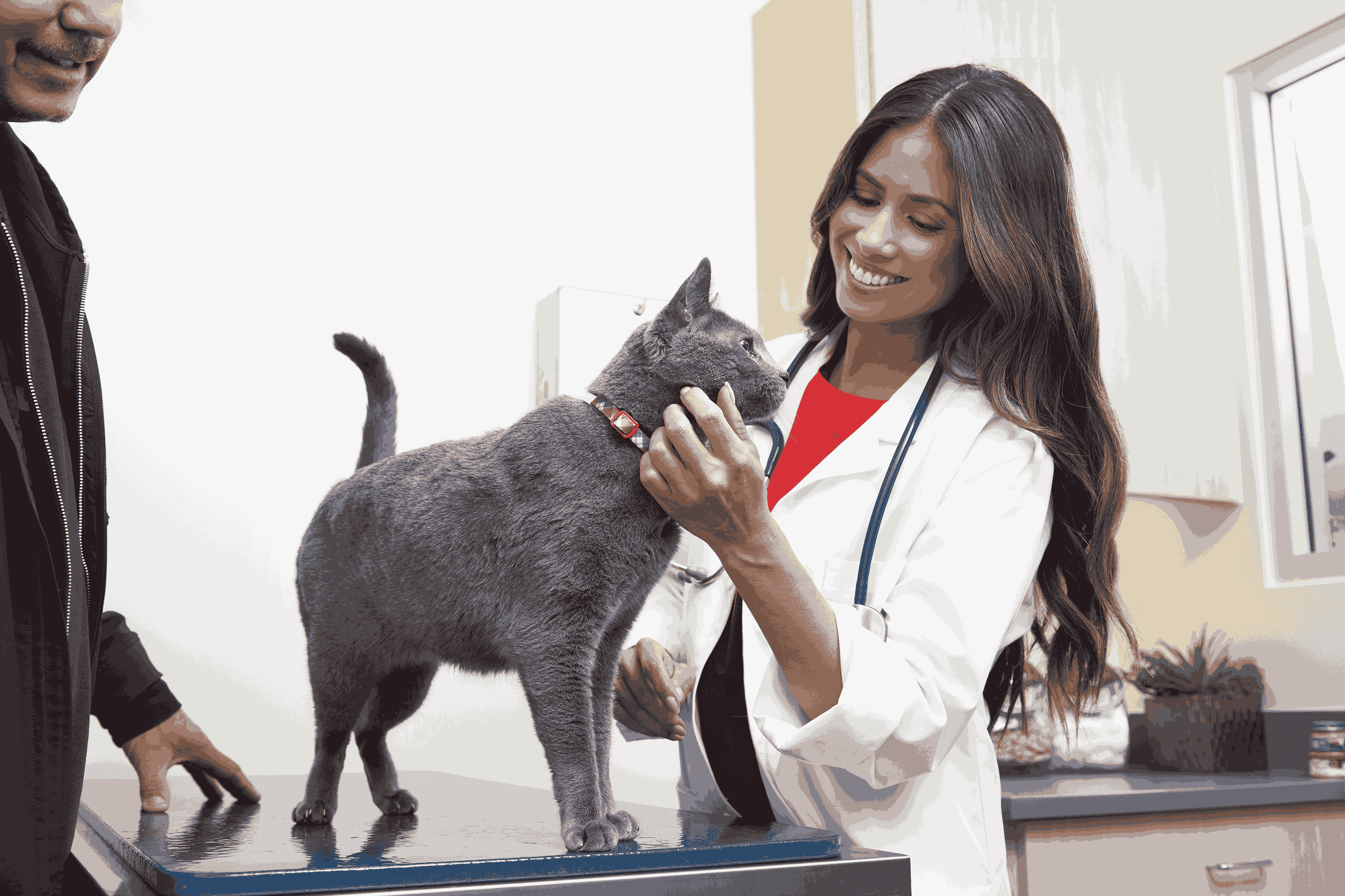
Diagnosis
Your veterinarian will begin with a physical examination of your dog. They will need to check for other causes of swelling in this area, which can include enlarged lymph nodes, tumors and abscesses. Your veterinarian will feel the enlarged area and take a sample with a needle to look at the cells under the microscope. This is called a fine needle aspirate. Based on the type of cells present, your veterinarian can likely determine what is causing the enlargement. If the cells are not clear, your veterinarian may recommend a biopsy, where a larger section of tissue is taken and sent to a lab. This may be done with full anesthesia or just sedation. This test is called a histopathology and can often confirm what is causing the enlargement. Your veterinarian may also recommend imaging, such as x-ray or CT scan to see how far the cysts extend so they can plan a surgical approach.
Causation
The salivary glands, as their name implies, are responsible for the production of saliva. Several sets of them lie in the tissue area beneath the skin in the upper and lower jaw. Saliva production goes on constantly. It continuously makes its way into the mouth through small ducts that originate within the salivary glands and course under the skin, opening directly into the oral cavity. The purpose of saliva is to lubricate the mouth, moisten the food, and begin the digestive process. Salivary cysts occur when saliva leaks from the salivary ducts into the tissues near or within the mouth. This may be caused simply by trauma to the ducts. In some instances, the duct may have been broken or bruised with the resultant swelling closing off the duct. In other cases, an abscess or tumor in the area may put enough outside pressure on the duct to pinch it off as you might a garden hose. Finally, there are instances when the glands, because of an internal inflammatory process, release a fluid that is just too thick to make its way through the tiny ducts. This again leads to an obstruction. All of these cases have the same final result. Whether the duct was originally ruptured or not, the pressure of continued saliva production by the glands will finally cause the walls of these tiny tubes to break and allow saliva to leak into the surrounding tissue. A small amount of a fluid leaking into tissue rarely, if ever, causes a problem and will usually go unnoticed. Saliva leaking into the tissues almost always develops into a major problem because the fluid is produced constantly and in large quantities. Additionally, possibly because of the viscosity, the body has a difficult time reabsorbing it. It simply continues to build and forms large cysts.
Treatments
AT-HOME CARE
SUPPORTIVE CARE
MEDICATIONS
DEVICES
SURGERY
SPECIALISTS
Cost Of Treatment
The cost of surgical treatment will vary widely based on location and level of expertise. At some clinics this may cost as much as $1000 or more depending on the size and location of the salivary mucocele.
Recovery
Most dogs recover well after surgery.
Monitoring
Your veterinarian will want to remove any sutures 10 to 14 days after surgery, and usually a full recheck exam is performed at this time.
Prevention
There is no known prevention for this condition.
Disclaimer
The information contained on this page is for educational purposes only. Treatment should only be provided under the advice of a veterinarian who has examined your pet under the laws applicable to your state of residence.


Have A Vet Question?
Book an appointment with the pros – our expert vets are here to help.